Acute Coronary Syndrome: Patho & Localizing for EMS
Case Study
I still remember the first interior STEMI I ran as a brand new paramedic. It was my second or third shift on my own, cleared and independent, in a primarily rural county with a moderate capability CIC about 45 minutes away. I’m not sure who was more stressed - me or my patient - when I hooked my syncope patient up to the monitor and ran my 12lead. The 12 lead ink smudged a bit on my glove as I read the massive inferior wall elevation with corresponding reciprocal changes in the lateral leads. Sure enough, I’d caught my first STEMI.
My equally clueless partner and I went to work. I missed my first line but got my second. The patient was moderately bradycardic at this point with a HR in the upper 40s and a BP in the 80s systolic. The patient kept clutching his chest. You’re giving me chest pain too, I thought to myself as I looked at him.
We hurriedly loaded him up and got him in the unit. My partner got up front and rocketed down the roadway, feeding off my own anxiety at that point. I called in my report as I gave him a fluid bolus - which thankfully relieved his hypotension. I kept wondering, why the hell is he bradycardic? Shouldn’t his stressed heart be tachycardic?
I walked into the ER feeling like a hero for my ever-so-valiant STEMI recognition, but the ER doc barely looked up at me during report and my patient was then shuttled rather unceremoniously away from me.
Introduction
Heart disease, and by extension acute coronary syndrome, is an epidemic in the United States. Since the 1950s, it has topped the charts of the leading causes of death and affects millions of Americans. While heart disease itself is an umbrella term, there are specific types of acute heart disease that EMS and the emergency room play outsized roles in ensuring survival and long-term health for. Our interventions during the acute phases of heart disease can make or break outcomes.
Acute Heart Disease Issues
In EMS and emergency medicine, we most commonly deal with acute coronary syndrome. As the name suggests, this is an acute disease of the coronary arteries that surround and feed the heart. Some pathology is disrupting blood flow and causing ischemia to the heart. Acute coronary syndrome itself is another umbrella term and includes ST Elevation Myocardial Infarction (STEMI), Non-ST Elevation Myocardial Infarction (NSTEMI), and Unstable Angina.
STEMI is a full, 100 percent occlusion of one of the coronary arteries. It is the most severe form of acute coronary syndrome with a 5 year survival rate (post-treatment) about 60 percent on average. This is a severe emergency that requires rapid, early intervention through percutaneous coronary intervention (PCI). If PCI is not readily available (for instance, in isolated rural emergency departments without the ability to quickly transfer patients out to a CIC), then fibrinolytics are the last resort treatment method. PCI involves directly stenting the coronaries involved to dislodge the clot and restore blood flow while fibrinolytics involve providing tPA/TNK to attempt to chemically clear the occlusion. Fibrinolytics have a lower success rate and are more risky than traditional PCI.
Symptoms of STEMI
In men, the classic symptoms of STEMI include crushing substernal chest pain with potential radiation to the left arm, jaw, or whole left side of the body. Accompanying symptoms include nausea/vomiting, dizziness, syncope/weakness (related to ischemia and compromised perfusion). Vision changes can also occur.
Women have more atypical STEMI presentations. Patients with comorbid conditions such as diabetes or previous cardiac injury also often present with atypical symptoms. For a female patient, sometimes the only symptom is unexplained weakness, nausea/vomiting, syncope, or even tooth pain.
While you should always maintain a high index of suspicion for all patients, it is critically important that we properly assess female patients and remember that their symptoms may not match males.
Other signs/symptoms include:
Physical gestures
A variety of named physical signs exist for ischemic heart disease, each with varying sensitivity. Classic examples include the Levine sign and the Palm Sign.
Diaphoresis & cool/clammy/pallorous skin
As catecholamines dump, the body begins to shunt blood to the central core and away from the periphery. This includes vasoconstriction of the small veins/venules and capillaries of the skin. This is not necessarily specific to ischemic heart disease.
Syncope
Related to cardiac perfusion changes. As the heart becomes more ischemic, cardiac output drops and the ability is less able to compensate for simple positional changes. Temporary disruptions in perfusion can cause syncope.
Diagnosis & Assessment
Most of the physical assessment findings that we see with STEMI (and NSTEMI) are related to changes in perfusion and the influx of catecholamines as a compensatory mechanism to ischemia.
STEMI is diagnosable through a combination of history/physical exam findings, troponins, and EKG assessment. Troponin is a type of structural protein only found within cardiac myocytes. During ischemia and infarct, troponins are released into the bloodstream and are measurable with lab testing. The amount of troponin present in lab draws is proportional to the degree of cardiac damage.
NSTEMI, as the name suggests, is a lesser occlusion without ST elevation present on EKGs. NSTEMI patients may have a normal 12lead or show some ischemic findings such as T wave inversion or ST depression but will not show ST elevation. Like STEMIs, NSTEMIs are diagnosed through physical exam findings/history and troponins; 12 leads have less utility.
Troponins are structural proteins found only within cardiac myocytes. As cells break down during myocardial damage (such as in ischemic heart disease), these proteins leak out into the plasma and become detectable with lab testing. It is important to remember that anything that does some form of damage to the heart can cause an elevation in troponin - for instance, blunt chest wall trauma of varying degree or sepsis can cause troponin to elevate. Sepsis patients have some form of elevated troponin in 85 percent of cases on average.
Localizing Coronary Arteries on EKGs
The precordial leads of a 12-lead EKG all offer specific views into different sections of the middle and left side of the heart. Unlike the traditional limb leads that make up aVR and leads I through III, precordial leads are strictly straight-ahead views of the electrical activity in front of them.
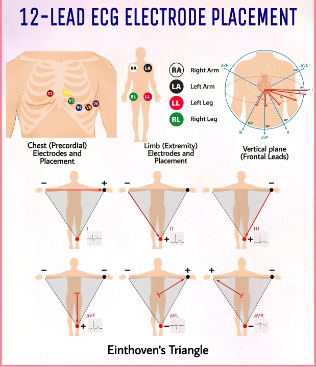
Three main coronary vessels perfuse the different sections of the heart. These coronary vessels branch into smaller vessels to cover each section and feed the heart muscle itself during diastole. We will cover each one in detail below, along with providing information that may help you predict symptoms early based on the EKG.
It is important to note that this article will only discuss the typical anatomy of the majority of the population. A significant percentage of the population has different vascularity with different coronary vessel placement.
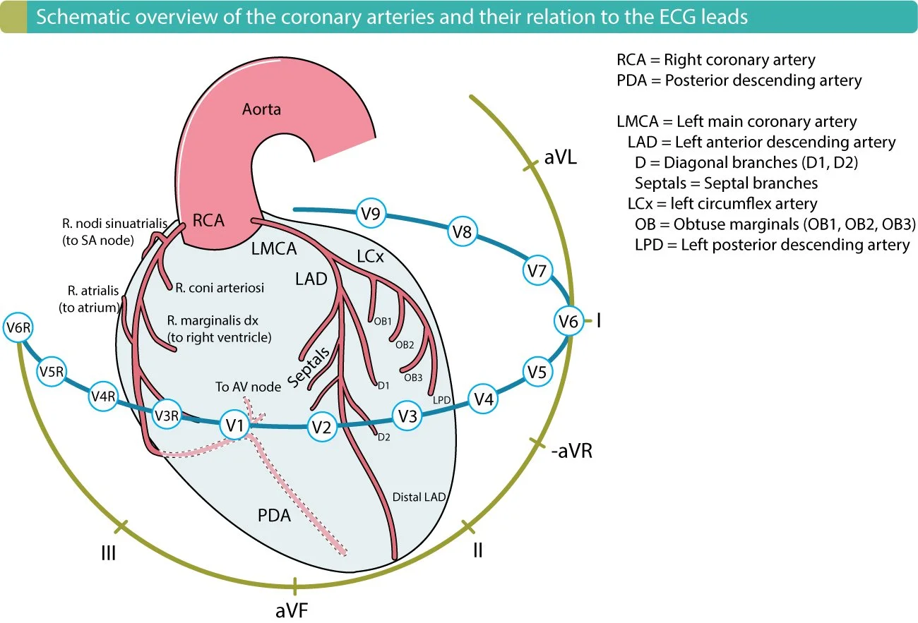
Left Main Coronary Artery
The left main is the largest coronary vessel and branches off from the left aortic sinus. It bifurcates into two large divisions; the left anterior descending and the left circumflex, which both primarily perfuse the left side of the heart including the left ventricle. Occlusions of the left main itself are catastrophic and typically result in rapid decline into cardiogenic shock and subsequent cardiac arrest. It is very common for these patients to not make it to the ER, and many of these patients unfortunately die in the field with unsuccessful prehospital resuscitations. Unlike most other STEMIs, left main coronary artery occlusions have somewhat better survival, if caught early, with surgical CABG management (Burgazli et al., 2012).
Left Anterior Descending (LAD) - The Widowmaker
The left anterior descending coronary artery branches off the left main and runs down to perfuse the anterior wall of the left side of the heart in addition to the septal wall. The LAD is massive compared to the other coronaries and carries over 50 percent of the total coronary vessel blood supply (Rehman et al., 2023).
As the LAD is occluded, damage occurs to the left ventricle and septal wall. The left ventricle is primarily responsible for maintaining cardiac output and is by far the most muscular portion of the heart. LAD occlusions The LV must be capable of opening the aortic valve to flow blood out into systemic circulation. Ejection fraction (EF) drops as the LV’s ability to pump is impaired.
Most of the symptoms associated with LAD occlusion are related to the accompanying drop in ejection fraction and cardiac output. Expect these patients to be acutely ill and be prepared to manage severe complications as they arise. Cardiogenic shock is common in these patients and may be irreversible.
Similarly, the septal wall is responsible for a significant portion of normal cardiac movement and ejection fraction. During systole, the septum twists and moves inwards towards the left ventricle to contribute pumping motion (Triposkiadis et al., 2022).
The Left Circumflex
The left circumflex artery branches off from the left main and mainly covers the lateral wall of the left side of the heart. It perfuses both the left ventricle and the left atria walls. Compromising the lateral wall’s ability to squeeze inwards can also significantly damage ejection fraction and the heart’s ability to pump.
Right Coronary Artery
The right coronary vessel primarily covers the right ventricle and right side of the heart. It is best represented by the inferior leads (II, III, aVF). It is less lethal than anterior STEMIs (LIFTL). While occlusions of the other coronary vessels typically result in classic cardiogenic shock, RCA occlusion specifically has some unique, prehospital-relevant ramifications.
The RCA provides perfusion to the right atrium and thus the sinotrial (SA) and atrioventricular (AV) nodes. The SA node is the natural pacemaker of the heart while the AV node plays a critical role in holding and transmitting impulses downwards into the ventricles to complete the cardiac cycle.
Inferior STEMI presents a high risk for bradycardia and AV block development as parts of the conduction pathway become ischemic. Even if the left ventricle and cardiac output are intact, the RCA-resulting symptomatic bradycardia can drop cardiac output on its own. While some degree of mild bradycardia with stable vitals is acceptable, we need to intervene if a bradyarrthyhmia is compromising perfusion.
Right ventricular involvement (RVMI) is also common with these patients at a rate of 10 to 50 percent (Jeffers et al., 2023). The right ventricular is a relatively weak, low-pressure part of the heart and is thus heavily dependent on preload to function. It does not have the muscularity to push against high pressure that the left ventricle does. Careful fluid resuscitation may be pertinent to help support preload.
The inferior leads do not directly look at the right ventricle. Switching to right-sided leads can help identify if the right ventricle is also experiencing infarct.
Traditional wisdom typically says that nitroglycerin and nitrates are contraindicated in the presence of RVMI. However, recent studies have found relatively low risk with few adverse events resulting from nitrate use (Wilkinson-Stokes et al., 2022). As with all things, use your best clinical judgement in the context of the patient’s overall condition.
Clinical pearls:
Avoid contributing to fluid overload and third spacing by being conservative with fluid resuscitation. Patients with a reduced EF do not respond well to increasing preload by adding more fluid volume. Remember that every drop of fluid you add is fluid that may need to be diuresed off later or may worsen cardiac stress in the short term.
Conclusion
The goal of this article is to expand on your knowledge about the underlying pathophysiology of acute coronary syndromes and the different areas of the heart that each lead represents. Use your best clinical judgement in dealing with these patients and, as with all things, follow your physician orders or standing orders in your area of practice.